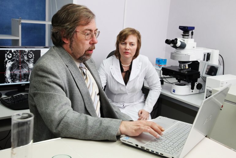
TSU neuroscientists are introducing a new diagnostic approach that will quantitatively evaluate myelin, the substance that makes up the sheath of nerve fibers. The scientists are searching for biomarkers of maturation and brain damage in children, including the fetus after 20 weeks of pregnancy. Their studies are aimed at reducing perinatal mortality and preventing serious diseases of the central nervous system and cognitive disorders, including autism.
- Congenital malformations of the fetus are among the three main causes of perinatal mortality and neonatal death,- says Vasily Yarnykh, Professor at the University of Washington (USA) and research supervisor of the TSU Laboratory of Neurobiology of the Research Institute of Biology and Biophysics. - Most of the anomalies are due to the interaction of genetic and environmental factors. The possibility of influencing them is still extremely limited, so today the main method of preventing serious diseases of the central nervous system is to create new tools for the early diagnosis of congenital anomalies and predict their outcome.
The TSU scientists suggest using mapping of myelin, one of the main components of the brain substance that conducts nerve impulses and protects nerve fibers from damage, to solve this problem.

The world's first noninvasive method of assessing myelin was developed several years ago under the guidance of Vasily Yarnykh, a scientist at TSU and the University of Washington. The new approach has already proven its effectiveness. Now it is being implemented at several clinics of the Russian Federation, including being used to assess damage to the sheaths of nerve fibers in people who have suffered a stroke. Under the new project, neurobiologists will use a unique approach to assess myelination in normal conditions and in different brain pathologies, in both the pre- and postnatal period of development.
- MRT in the prenatal period is an extremely difficult task because it is impossible to fix the fetus at the time of scanning. To obtain an objective picture, special programs are used to combine images, - explains Marina Khodanovich, head of the Laboratory of Neurobiology at the TSU Research Institute of Biology and Biophysics. – Research is planned at the International Tomographic Center of the Siberian Branch of the Russian Academy of Sciences (Novosibirsk) and will be conducted under the supervision of Dr. Alexandra Korostyshevskaya, who is a pioneer in the use of prenatal MRT in Russia.
In 2018, the new method was already tested when examining children in the first years of life and fetuses in the process of intrauterine development. Scientists obtained new data on the features of the development of human brain structures during their intrauterine development. It has been established that the amount of myelin in the fetus in the second trimester is minimal; first of all, it is formed in the structures that are responsible for vital functions - respiration, blood circulation, and heart activity. In the third trimester, the initial myelination of the structures responsible for motor functions and coordination of movements becomes noticeable. During the examination of children 2-3 years old, a number of cases were revealed when the new method obtained additional diagnostic information useful for doctors.
Research supported by the Russian National Science Foundation will be conducted over four years. It will optimize the technology of quantitative myelin mapping at the pre- and postnatal stages and to create a standard quantitative digital atlas of myelination of the fetus and child's brain.

The data will help to open a new way to determine the causes of delayed development in children and predict their intellectual abilities. Diagnosing abnormalities of myelination at an early age is also important in identifying risk groups of patients for mental retardation, autism, and more serious mental illnesses, which in turn can be used to prescribe early behavioral therapy and develop social adaptation skills.
