Not only is it difficult to diagnose and pinpoint an early-stage brain tumor—certain difficulties may arise even after the onset of symptoms. Tomsk State University scientists have developed a new noninvasive approach to diagnosing a glioma, one of the most severe types of brain tumor. Using Raman spectroscopy, they detected biotraces—chemical compounds secreted by a tumor—in blood. The research is presented in an article published in Pharmaceutics (MDPI, Q1).
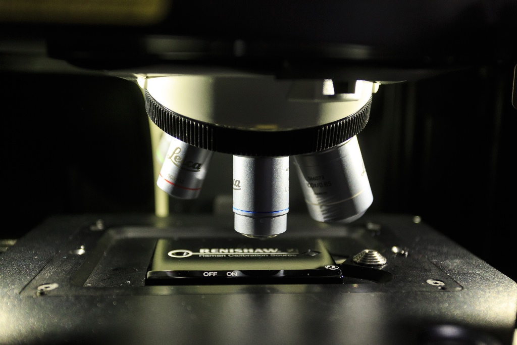
“It is possible to verify the type of a tumor only through histological examination, which is carried out after the neoplasm has been removed,” comments Yury Kistenev, head of the TSU Laboratory of Laser Molecular Imaging and Machine Learning. “However, optical analysis methods significantly expand the diagnostic possibilities and, moreover, enable us to do everything noninvasively, without taking biological tissues. That being said, we used Raman spectroscopy, which helps detect chemical compounds in biological fluids and tissues with high accuracy.”
At the initial stage, scientists worked with the field-specific literature and collected information on the types of biological markers that are found in glioma tissues. They then conducted an experiment on laboratory animals: Mice were injected with human glioblastoma cells, and after different periods of time their blood serum was examined, and they themselves were removed from the experiment. By means of Raman spectroscopy, scientists identified the specific frequencies that make it possible to pinpoint traces of glioma in biological fluids and divide the mice into the healthy and unhealthy groups.
New data was collected into a library that the research team used to train a neural network that helped automate the analysis process.
“What makes our approach advantageous is that it enables us to find traces of an early-stage tumor, prior to symptoms manifesting themselves,” explains Yury Kistenev. “The thing is, a glioblastoma changes the biochemical composition of blood. It secretes substances and tumor cells that end up in blood, which carries them around the whole body. By analyzing these biotraces we can get data on cancer formation: That information in turn can be used to diagnosing a disease or monitoring the effectiveness of treatment.
According to the scientists, the new approach may prove to be promising for detecting other types of tumors. All malignant neoplasms secrete specific chemical compounds, and if it is known which exact biotraces are related to a particular medical condition, they can be detected using Raman spectroscopy and machine learning.
Optical analysis methods are becoming more common in various fields; in particular, they are opening up great opportunities for quick diagnosis of diseases. In this regard, supported by a megagrant of the government of the Russian Federation, TSU scientists are using AI to develop new noninvasive methods of diagnosing viral and bacterial respiratory infections, which will reduce the time needed for analysis from several days to several minutes.